FHWN 0519 4888 Pletterbauer

Biomed Research & Image-guided Therapy

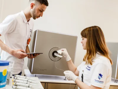
The main focus comprises the quantification and visualization of anatomical and structural information, as well as complex biochemical processes at the cellular and molecular level in living cells, biological and artificial tissues and in the living organism using morphological, functional and molecular imaging techniques, including the development and evaluation of novel imaging biomarkers and methods for specific (patho)physiological processes.

Another focus is on the carrying out of laboratory analytical tests as part of the biomedical analysis process. The main focus here is on (immuno)histological investigations of a wide variety of tissue samples, including digitalization of the results as well as the performance of titer studies.

We perform research and development in the following topics:

  • Development and evaluation of imaging techniques to facilitate the understanding of the systems biology of different tumor entities
  • Visualization and quantification of highly specific tumor(patho)physiological properties using radiolabeled biomarkers and their combination with multimodal preclinical morphological, functional and molecular imaging techniques (micro-CT, micro-SPECT, micro-PET and micro-MRI)
  • Application, establishment and validation of preclinical molecular and semi-functional imaging methods in radiotherapy and their use in applied research in the field of biomedicine, radiobiology and medical radiation physics
  • Investigation of complex biological systems and bioinformatic data analysis
  • Investigation of imaging biomarkers and the effect of drugs at the molecular level
  • NGS data analysis in personalized medicine (genomics, metagenomics and transcriptomics)

Infrastructure:

  • micro-Computed Tomography (X-Cube, Molecubes)
  • micro-Positron Emission Tomography (b-Cube, Molecubes)
  • micro-Single Photon Emission Tomography (g-Cube, Molecubes)
  • micro-Magnetic Resonance Tomography (15.2T UHF, Bruker BioSpin)
  • Biomedical image data analysis, reconstruction and complex intelligent systems
  • Image Post-Processing Laboratory
  • Small Animal Housing Unit
  • Ultrasound Laboratory
  • Radiotherapy Laboratory
  • Nuclear Medicine Laboratory
  • Intervention Laboratory
  • X-ray and Mammography Laboratory
  • Biomedical Analytics Training Laboratories
  • LC-HRMS
  • Nano LC-MS/MS
  • HPLC
  • GC-MS

Research Activities

  • Ra-Dia_M

    In the field of cancer diagnostics, the IMC Krems University of Applied Sciences and the Wiener Neustadt Biotech Campus Tulln University of Applied Sciences are jointly breaking new ground in Lower Austria. The effectiveness of drugs on cancer cells is examined under the so-called Raman microscope and evaluated with artificial intelligence (AI).

  • PAIR

    Cancer therapy has experienced a rapid surge in the use of protons and carbon ions, over high-energy photons. This is due to their superior physical properties, including their major energy deposition at the end of their range and their higher ionization densities of particle beams. The biological outcomes are less damage to healthy tissue, as well as altered DNA damage and cellular signaling.

  • PRECISE

    Given the importance of successful implantation procedures, the study of osseointegration and the factors influencing this process is a leading topic in orthopedics and dentistry. The rat tibia implantation model is a widespread animal test that evaluates the osseointegration of implants under pre-clinical conditions in vivo, for instance, to determine whether a newly developed implant meets compatibility, safety, and mechanical stability requirements. This proposal follows the hypothesis that the application of tailored surgical guides improves the accuracy, precision, and reproducibility of implant positioning, as well as the overall performance of the rat tibia implantation model, thereby resulting in the refinement and reduction of animal testing for osseointegration studies.

  • Angewandte molekulare Bildgebung

    The current project aimed to foster the understanding of the systems biology of a various number of different tumor entities, increasing the knowledge about radiobiological aspects and help to facilitate the integration in the field of radiation therapy planning, verification and treatment monitoring. In this context, the planned R&D research infrastructure will be implemented in the research facilities of the University of Applied Sciences Wiener Neustadt and the ion therapy and research center – MedAustron to support (1) expert training of students and scientific staff in theoretical (lectures) and practical (“hands-on training) aspects of preclinical molecular imaging techniques, (2) collaborative research especially in the field of functional imaging, medical physics and radiation biology to investigate molecular changes in tumor biology and microenvironment during radiotherapy, (3) acquisition, performance analysis and verification of preclinical imaging techniques in radiation therapy. The intention of the R&D research infrastructure is to provide the basis for cutting-edge biomedical research in the field of both conventional and ion radiotherapy in a regional, national and international context. In addition the project benefits and increase the quality of the education in the academic environment (students, scientific staff, etc…) in the field of quantitative molecular imaging, medical physics, radiobiology, radiation therapy planning and verification and helps to increase the understanding of (patho)physiological processes concerning the systems biology of a various number of different tumor entities.

Banner

Contact

Feel free to contact us: FuE@fhwn.ac.at